
Eye reconstruction and ocular prosthesis
Reconstruction of the eye and the use of ocular prostheses are important aspects of oculoplastic surgery and ophthalmic surgery.
The eye can be irreversibly damaged by serious illnesses (trauma, infection, tumor, complicated glaucoma). When preservation of the eye is not possible, evisceration or enucleation may be performed, followed by placement of an ocular prosthesis to restore the natural appearance of the eye.
The eye must then be replaced by a prosthesis in the orbit called a ball which will be covered and integrated by the tissues around the eye.
We call this
- a evisceration
- an enucleation
- or an exenteration.
Eye reconstruction aims to restore the appearance of the eye: it is an ocular prosthesis made by an oculoprosthetist to restore the natural appearance of the eye.
The ocular prosthesis is adapted to the shape of the orbit and is custom-made for each individual.
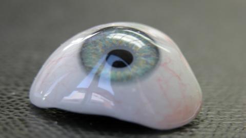
Prosthetic eyes are designed to resemble a natural eye and match the shape and color of the iris of the other eye. They are made from safe and biocompatible materials such as resin or glass. They are individually adapted by a specialist in ocular prostheses, called an ocularist. They should be maintained regularly to ensure proper hygiene and a comfortable fit. However, they do not allow vision. They are designed for aesthetic reasons only.
Specialized ocularists work closely with ophthalmologists in Geneva to provide high-quality, personalized ocular prostheses.
Over the years, certain complications can arise: loss of fat volume, rejection of the ball, instability of the prosthesis. This requires reconstruction of the orbital cavity which is most often surgical:
- dermofat graft
- oral mucosa graft
- lipostructure with lipofilling
evisceration
Evisceration is an eye surgery during which the contents of the eye are removed but the outer covering of the eye called the sclera is retained as well as the eye muscles attached to it. This eye surgery is unfortunately necessary in the case of total loss of vision due to a painful eye despite all non-invasive treatments.
It can also be justified for aesthetic and social reasons because an eye that is no longer viable atrophies and becomes white, which is called ocular phthysis.
The stages of this surgery are as follows: firstly, local anesthesia is performed by an injection behind the eye in order to eliminate any pain related to the procedure. General anesthesia may also be given. The procedure is then carried out to remove the ocular contents but the optic nerve is preserved in cases of evisceration.
Once this step has been completed, an ocular implant or “ball” is placed inside the scleral volume after measuring its contents. This ball is made of a biocolonizable material to ensure its integration by the tissues of the orbit. Then the sclera, tenon and conjunctiva are sutured plane by plane.
Finally, a temporary conformator is positioned in the conjunctival cul-de-sac while awaiting the creation of an ocular prosthesis by the ocularist six weeks later.
Here is some information about this surgery:
1h30 with overnight hospitalisation
general
- Anesthesia consultation before surgery
- Be fasting on the day of the operation
- Exit the day after the procedure with a bandage on the eye
- Do not play sports or put your head under water for a week
- Antibiotic therapy will be prescribed for one week
- Care is provided at home to redo the dressing daily for a week
enucleation
Enucleation is an eye surgery very similar to evisceration. The main difference being the section of the optic nerve in enucleation, absent in evisceration. Thus, this surgery allows when necessary to remove the entire eye, that is to say the entire eyeball, when the scleral part cannot be preserved. These are mainly cases of malignant intraocular tumor, serious trauma, severe infection or ocular pain resistant to any treatment.
The stages of the surgery are as follows: First, general anesthesia is given. The procedure is then carried out to remove the entire eye after sectioning the optic nerve. During this procedure the eye muscles attached to the sclera are removed and preserved. After having carried out a measurement of the orbital cavity, an implant covered and chosen according to the appropriate size on which the oculomotor muscles are sutured. The tissues are then closed plane by plane and a conformator is positioned in the cavity while awaiting the manufacture of an ocular prosthesis by the ocularist six weeks later.
In some cases enucleation is carried out but the sclera is cleaned and preserved in order to cover the ball or implant positioned in the orbital cavity.
Here is some information about this surgery:
1h30 with overnight hospitalisation
general
- Anesthesia consultation before surgery
- Be fasting on the day of the operation
- Exit the day after the procedure with a bandage on the eye
- Do not play sports or put your head under water for a week
- Antibiotic therapy will be prescribed for one week
- Care is provided at home to redo the dressing daily for a week
Day 1: appointment with Dr Bela for monitoring
Day 7: dressing removed
M1: a temporary prosthesis is made
M6: Once the wound has healed completely, the ocular prosthesis specialist makes a definitive made-to-measure prosthesis. This requires no handling on the part of the patient and all activities are possible. The prosthesis is polished every 6 months and replaced every 5 years.
dermal graft
Dermofat grafting is a surgery performed by the oculoplastic surgeon. This is an autologous tissue graft, that is to say specific to the patient, composed of dermis and fat from a part of the body in order to transplant it to an area requiring filling following a loss of volume.
In the orbit, these are mainly cases where the ball acting as a prosthesis in the orbital cavity is not tolerated by the patient. This rejection requires removal of the ball, making the orbital cavity empty and hollow. In order to allow adaptation of a new ocular prosthesis it is necessary to provide a volume which compensates for the removed ball in order to allow support of the ocular prosthesis.
This dermofat graft is most often taken from the abdomen and then this graft is carefully placed in the eye cavity and sutured to the oculomotor muscles if possible.
Finally, the superficial tissues are sutured layer by layer and an ocular shaper is positioned in the cavity while awaiting the creation of a new ocular prosthesis by the ocularist six weeks later.
- The advantage is that there is no risk of rejection of the dermofat graft.
- The disadvantage of a dermofat graft compared to evisceration or enucleation is the unpredictable volume of the residual graft as well as the virtual absence of ocular movement once the prosthesis has been adapted into the cavity. This has essentially aesthetic consequences.
Here is some information about this surgery:
1h30 with overnight hospitalisation
general
- Anesthesia consultation before surgery
- Be fasting on the day of the operation
- Exit the day after the procedure with a bandage on the eye
- Ne pas faire de sport ni mettre la tête sous l’eau pendant une semaine
- Une antibiothérapie sera prescrite pour une semaine
- Des soins sont réalisés à domicile pour refaire le pansement quotidiennement pendant une semaine
Day 1: appointment with Dr Bela for monitoring
Day 7: dressing removed
M1: a temporary prosthesis is made
M6: Once the wound has healed completely, the ocular prosthesis specialist makes a definitive made-to-measure prosthesis. This requires no handling on the part of the patient and all activities are possible. The prosthesis is polished every 6 months and replaced every 5 years.
Oral mucosa grafT
Oral mucosa grafting is a surgery performed to replace tissue loss in the orbital cavity.
This is often carried out following evisceration or enucleation when there is a loss or insufficient volume of the conjunctiva. It may also be justified in cases of scar retraction of the cul-de-sacs linked to a conjunctival or post-traumatic fibrosing disease. When the cavity lacks volume at the level of the conjunctiva, this creates retracted or too short dead ends making the manufacture and adaptation of the ocular prosthesis difficult for the ocularist.
When there is fibrosis of the flanges or symblepharons on the conjunctiva, this can cause retraction of the eyelid and eyelashes, resulting in what is called scar entropion. This condition on a seeing eye can cause serious damage due to the daily friction of the eyelashes on the cornea, ultimately causing loss of vision with a poor visual prognosis.
The oral mucosa is a very thin tissue, taken from the inner surface of the lip. This tissue has anatomical and structural components similar to that of the conjunctiva.
This intervention therefore makes it possible to carry out a graft in order to restore the missing volumes. This will make it possible to restore the orbital cavity which will accommodate the definitive prosthesis or to restore the dynamics of the eyelids and the orientation of the eyelashes to protect the eyeball.
Here is some information about this surgery:
1h30 with overnight hospitalisation
general
- Anesthesia consultation before surgery
- Be fasting on the day of the operation
- Exit the day after the operation with a bandage on the eye. No sutures in the mouth
- Do not play sports or put your head under water for a week
- Antibiotic therapy will be prescribed for one week
- Liquid and cold food during the first week.
- Care is provided at home to redo the dressing daily for a week
D1: Appointment with Dr. Bela for monitoring
D7: removal of the dressing
M1: After complete healing, the ocular prosthetist adjusts a final custom prosthesis. This does not require manipulation on the part of the patient and all activities are possible. Polishing by the prosthetist is carried out every 6 months and a change every 5 years.
Lipostructure
The lipostructure can be carried out for aesthetic purposes in order to fill a lack of volume but also in the case of orbital cavity surgeries, the lipostructure is carried out for a functional purpose.
Indeed, the adaptation of a glass or resin ocular prosthesis following eye evisceration or enucleation surgery requires a sufficient volume of support in the orbital cavity.
Over time, atrophy develops of the orbital tissues and the vessels that supply the eye before excision surgery. This atrophy is mainly linked to a loss of orbital fat volume. This causes a lack of support for the ocular prosthesis which will gradually move downwards and forwards. This has an aesthetic consequence because the upper eyelid deepens, the prosthesis moves, and the projection of the pupil is no longer the same as that of the contralateral seeing eye.
The treatment of this syndrome called “enucleum syndrome” consists of carrying out a transplant of adipose tissue taken from the patient.
This fat is therefore collected using a fine cannula, either from the inside of the knees, from the hips or from the abdomen. Then it is rinsed, filtered or centrifuged and prepared to be reinjected into the eye cavity, around the eyelids and eyebrow in order to restore lost volumes and symmetrize the look with the contralateral side.
After three months, the final volume of this fat is reached and allows the creation of a new prosthesis in a restored cavity.
This surgery lasts several years and can be repeated if necessary in the event of further loss of volume.
Here is some information about this surgery:
1h30 with overnight hospitalisation
general
- Consultation d’anesthésie avant la chirurgie
- Etre à jeun le jour de l’opération
- Sortie de lendemain de l’intervention avec un pansement sur l’oeil.
- Do not play sports or put your head under water for a week
- Antibiotic therapy will be prescribed for one week
- Care is provided at home to redo the dressing daily for a week
D1: Appointment with Dr. Bela for monitoring
D7: removal of the dressing
M3: The final results are visible after 3 months because this fat decreases by 20% within 3 months. After complete healing, the ocular prosthetist adjusts the custom-made prosthesis. This does not require manipulation on the part of the patient and all activities are possible. Polishing by the prosthetist is carried out every 6 months and a change every 5 years.
Can you see with an ocular prosthesis?
No, an ocular prosthesis, also known as a glass eye, does not allow vision. An ocular prosthesis is designed to replace the appearance of the natural eye after evisceration or enucleation surgery. It is made to measure by an ocular prosthetist or ocularist to recreate the exact volume of the eye in the socket, the exact colour of the iris and to allow eye movement. However, vision is no longer possible following the operation, so its purpose is purely aesthetic.
How does an ocular prosthesis work?
The ocular prosthesis is made by an ocularist. The ocularist takes measurements of the conjunctival cul-de-sac, known as an impression. From this impression, the ocularist will proceed to make a custom-made prosthesis by hand to recreate the exact colour of the iris and position this implant in the ocular cavity. The ocular prosthesis is often mistakenly called a "glass eye", but in reality it is made of resin, which is better tolerated than glass. Its purpose is to replace the appearance of the eye that has been lost through disease. The ocular prosthesis can be very thin (called a scleral lens) and positioned over a non-seeing eye that is present. Its purpose is purely aesthetic. More often than not, the ocular prosthesis is thicker than a scleral lens and is used both to fit an aesthetic ocular prosthesis and to restore volume in order to project the eye and support the eyelids following evisceration or enucleation surgery. Dr Bela works with a renowned ocularist based in Geneva, who can produce custom-made prostheses in just one day.
How often should I replace my ocular prosthesis?
It depends on the material of the prosthesis. Resin ocular prostheses are more resistant and better tolerated than glass prostheses. Apart from daily lubrication of the eye, the prosthesis will be checked and polished by the ocularist once a year. If discomfort is experienced, grinding may be used to adjust the volume of the prosthesis in the cavity. Because of the natural wear and tear of the materials, the prosthesis will be replaced every five years on average.
Is it possible to transplant an eye?
At present, science does not allow us to transplant an eye in order to restore vision that has been lost. However, science is constantly evolving, which is why an operation such as evisceration or enucleation is only reserved for cases where the eye is absolutely and totally blind. This corresponds to a total absence of light perception. In the case of a purely aesthetic problem in an eye that is not sighted but can perceive light, it is preferable to fit a thin, purely aesthetic prosthesis, known as a scleral lens, over the eye.